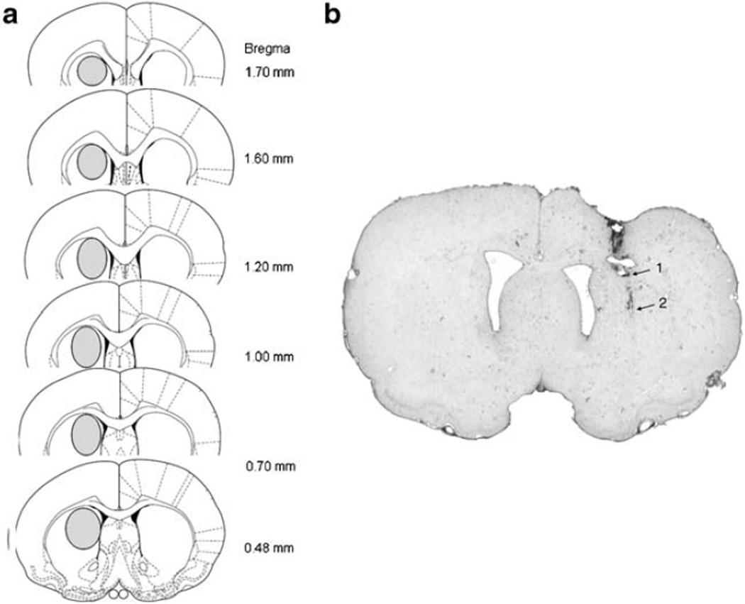
Fig. 1.
Location of microdialysis probes in the DSTR. a The areas of the DSTR where the probes were located are shown with an oval in each brain section. b Photo of representative probe placement in the DSTR. Arrow 1 indicates the end of the probe shaft and arrow 2 indicates the location of the tip of the probe membrane

