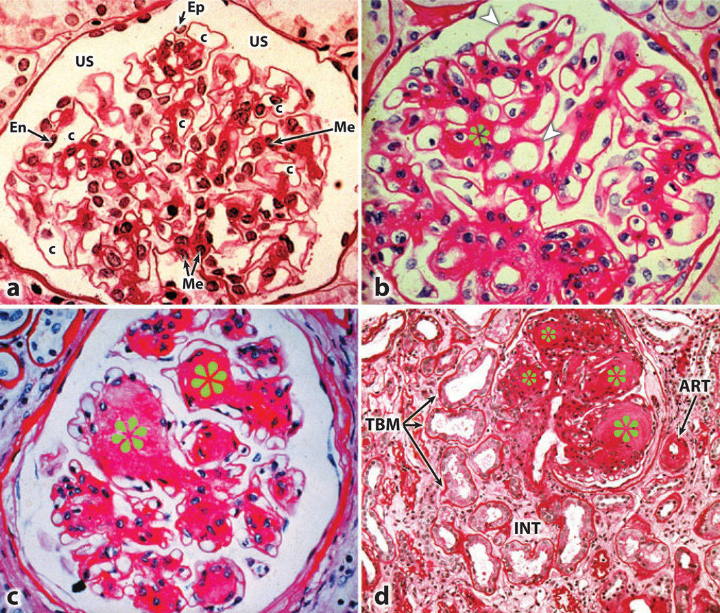
Figure 1.
Light photomicrographs illustrating various stages of developing glomerular lesions and tubulointerstitial disease in diabetic nephropathy. (a) A normal glomerulus. (b) Thickened basement membranes (arrowheads) and expanded mesangial regions (asterisks). (c) The nodular appearance of the mesangial regions characteristic of Kimmelstiel-Wilson lesions (asterisks). (d) The tubulointerstitial lesions include thickened tubular basement membrane (TBM), hyalinization of afferent arteriole (ART), and fibrosis of the interstitium (INT). Abbreviations: c, capillary lumen; En, endothelial cell; Ep, visceral epithelial cell (podocyte); Me, mesangium; US, urinary space.

