Figure 3.
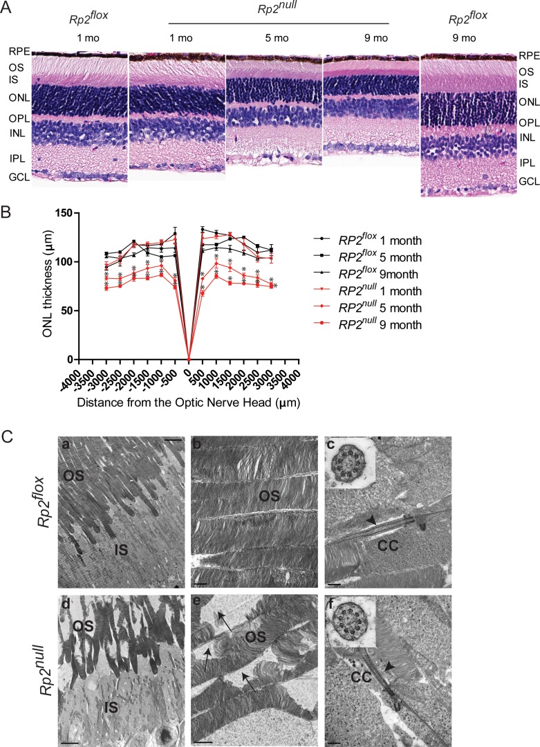
Retinal histology and TEM of Rp2null retina. (A) Histologic analysis of paraffin sections of Rp2flox (control) or Rp2null mouse retina was performed at indicated ages. RPE, retinal pigment epithelium; OS, outer segment; IS, inner segment; ONL, outer nuclear layer; OPL, outer plexiform layer; INL, inner nuclear layer; IPL, inner plexiform layer; GCL, ganglion cell layer. (B) Morphometric analysis of ONL thickness of 1, 5, and 9 months Rp2flox and Rp2null retinas (n = 5). The ONL thickness was measured in μm along the vertical meridian at each defined distance from the optic nerve head. Data represent mean ± SEM. *P < 0.01. (C) TEM analysis of control (Rp2flox, top) and mutant (Rp2null, bottom) photoreceptors at 10 months of age show disorganized OS in the mutant retina. Insets (c, f) show cross-sectional view of the connecting cilium (CC) with 9 + 0 array of microtubules in both control and mutant mice. Arrows in (e) indicate degenerated OS discs in the mutant retina; arrowheads in (c, f) show normal morphology of the CC. Scale: (a, d) 5.0 μm; (b, c, f) 0.5 μm; (e): 1 μm.

