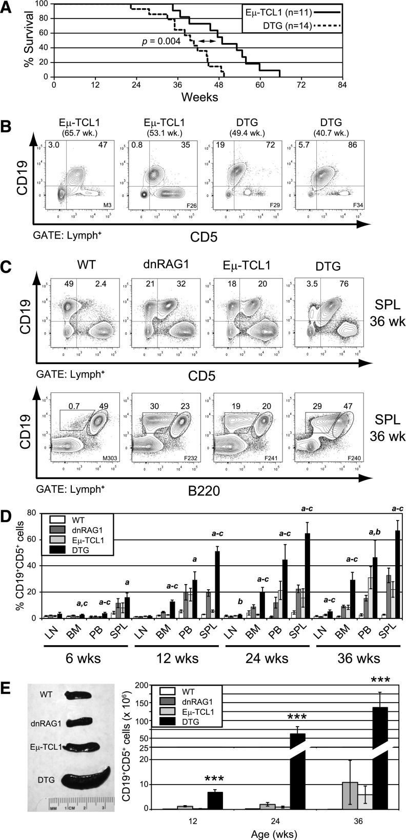
Figure 1.
dnRAG1 expression in Eμ-TCL1 mice shortens lifespan and accelerates CD5+ B-cell accumulation. (A) Kaplan-Meier survival curves of Eμ-TCL1 (n = 11) and DTG (n = 14) mice. Statistical analysis between groups was performed using the log-rank test (median survival: 39.6 and 49.0 weeks for DTG and Eμ-TCL1 mice; P = .004). (B) The percentage of splenic CD19+CD5+ B cells among gated lymphocytes is shown at end point for 2 representative Eμ-TCL1 and DTG mice. (C) Flow cytometric analysis of splenic lymphocytes from 36-week-old WT, dnRAG1, Eμ-TCL1, and DTG mice showing the expression of CD19 and either CD5 (top row) or B220 (bottom row). The percentage of CD19+CD5+, CD19+CD5−, CD19+B220hi, CD19+B220lo cells is shown for representative animals. (D) The percentage of CD19+CD5+ cells in lymph node (LN), bone marrow (BM), peripheral blood (PB), and spleen (SPL) of 6-, 12-, 24-, and 36-week-old WT, dnRAG1, Eμ-TCL1, and DTG mice (n = 5-6 animals per genotype per time point) was determined using flow cytometry as in panel A and presented in bar graph format. Error bars represent the SEM. Statistically significant differences (P < .05) between values obtained for DTG mice relative to WT (a), dnRAG1 (b), or Eμ-TCL1 (c) mice are indicated. (E) Spleen cellularities were measured at 12, 24, and 36 weeks of age for WT, dnRAG1, Eμ-TCL1, and DTG to determine the frequency of splenic CD19+CD5+ cells. Spleens from representative animals of each genotype are shown at left, and the frequency of CD19+CD5+ cells for each genotype and time point are presented in bar graph format at right. Error bars represent the SEM. ***Values obtained for DTG mice are significantly different (P < .001) from those obtained for WT, dnRAG1, or Eμ-TCL1 mice.

