
Fig. 4. γ-Aminobutyric acid (GABA) induced divergent Ca2+ responses in SCN neurons.
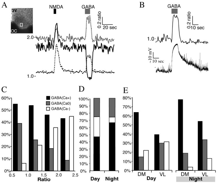
(A) SCN neurons imaged during the day, and treated with N-methyl-d-aspartate (NMDA; 200 μm, 5 s), which induced an increase in the Ca2+ ratio in all neurons. GABA (200 μm, 10 s) application induced divergent Ca2+ responses. The box within the SCN indicates the region recorded. Note the range of baseline Ca2+ concentrations. (B) Example of a GABA-induced rise in Ca2+ ratio and membrane depolarization in a SCN neuron recorded during the night using a loose-seal recording electrode. (C) The Ca2+ response to GABA (200 μm, 10 s) application varied with the baseline Ca2+ ratio. Ca2+ responses were separated into three groups: a transient elevation in the Ca2+ ratio [GABA(Ca+)]; a decrease in the Ca2+ ratio [GABA(Ca-)]; or no change in the Ca2+ ratio [GABA(Ca0)]. Data are the percentage of neurons in each response group based on the baseline Ca2+ ratio (range 0.5-1.0, n = 257 neurons; 1.0-1.5, n = 395; 1.5-2.0, n = 306; 2.0-2.5, n = 56). Note that the percentages of GABA(Ca-) and GABA(Ca0) neurons appeared reversed between low and high baseline Ca2+ ratios. (D) The percentage in the SCN neuronal response to GABA (200 μm, 10 s) varied between the day (n = 760) and night (n = 292). Note the change in the GABA(Ca+) and GABA(Ca-) groups, but not in the percentage of GABA(Ca0) neurons responding. (E) The percentage of SCN neurons with GABA-induced changes in the Ca2+ ratio varied between the dorsomedial (DM; n = 226 day and 145 night neurons) and ventrolateral (VL; n = 534 day and 147 night) regions of the SCN. The percentage of GABA(Ca+) neurons was higher in the DM during both the day and night. Conversely, the percentage of GABA(Ca-) and GABA(Ca0) neurons was higher in the VL in both day and night. 3V, 3rd ventricle; OC, optic chiasm.