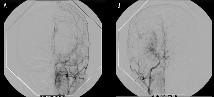
Figures 3.
(A) Left common carotid angiography showing extensive external carotid lepto-meningeal collateralization (occipital artery-ethmoidal branch of maxillary division-middle meningeal-anterior falcine). There is complete occlusion of internal carotid proximal to ophthalmic segment. (B) Right common carotid angiography showing attenuation of internal carotid with developed deep lenticulo-striate vessels as well as lepto-meningeal collaterals from external carotid. There is also crossover flow from right to left into the contralateral anterior cerebral artery.

