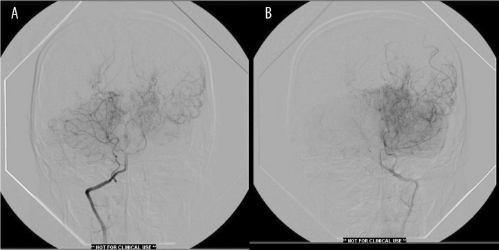
Figure 4.
(A) Right vertebral angiography showing extracranial origin of PICA. There is patent left posterior communicating artery with flow into middle cerebral artery distribution and evidence of choroidal vessel hypertrophy. (B) Left vertebral angiography showing typical findings with vertebral and posterior cerebral arteries poorly visualized.

