
Fig 1. PD-1 blockade in vivo was associated with increased Foxp3 expression and decreased PD-1 expression in CD4+CD25+ T cells.
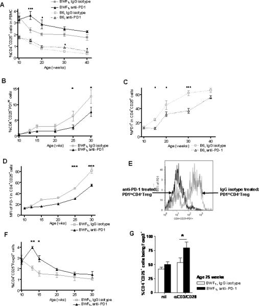
(A) PD-1 blockade attenuated the decrease in percentage of CD4+CD25+ cells with age. At a young age (10 week old) of BWF1 and B6 mice, in vivo PD-1 blockade maintained the percent of CD4+Treg at a higher level than in control mice (BWF1 and B6, p < 0.01, two-way ANOVA). (B) Percentage of PD-1hi CD4+CD25+ cells went up with age in BWF1 mice, but anti-PD-1 treated mice expressed fewer PD1hiCD4+Treg when compared to control mice. (C) Fewer cells expressed PD-1 in B6 compared to BWF1 mice. (D) MFI for PD-1 expression in CD4+CD25+ cells was higher in BWF1 mice treated with anti-PD-1 in vivo than in mice treated with IgG isotype (Fig. 1 A-D, p < 0.01, two-way ANOVA). (E) Representative histogram of the MFI for PD-1 in CD4+CD25+PD1+ cells from an anti-PD1-treated BWF1 mouse vs. an IgG isotype-treated control (at age 16 weeks) showing that PD-1 expression was reduced by treatment with anti-PD-1 Ab. We referred hereafter to the PD1+CD4+Treg derived from (in vivo) anti-PD-1 treated mice as PD1loCD4+Treg, and those from IgG isotype controls as PD1hiCD4+Treg. (F) Expression of Foxp3 in CD4+CD25+ cells went down with age in BWF1 mice, but anti-PD-1 treated mice had a higher Foxp3 expression than control mice until after age 15 weeks (Fig. 1F, p < 0.01, two-way ANOVA). (G) There was significant increase in the percent of CD4+CD25+Foxp3+Treg from mouse spleens at age 22 weeks after in vivo PD-1 blockade upon stimulation of TCR (p <0.05, unpaired student t-test). * p <0.05, ** p <0.01 (Bonferroni posttest). B6, n = 6 mice per group, BWF1, n = 14 mice per group.