
Fig 2. Inhibition of PD-1 in vivo increased the regulatory capacity of BWF1 CD4+Treg in different spleen cell subsets.
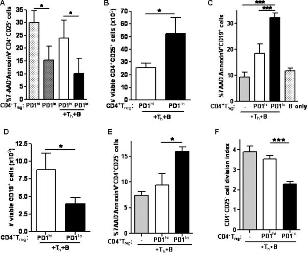
(A) Apoptosis in PD1loCD4+Treg was significantly less compared to PD1hiCD4+Treg, whether or not cultures contained Th and B cells (p < 0.05, unpaired student t-test). (B) PD1loCD4+Treg were viable and were increased in numbers compared to PD1hiCD4+Treg (p < 0.05, unpaired student t-test). (C) PD1loCD4+Treg induced more apoptosis in CD19+ B cells than did PD1hiCD4+Treg. (D) Numbers of viable B cells were decreased in cultures containing PD1loCD4+Treg compared to those with PD1hiCD4+Treg (p < 0.05, unpaired student t-test). (E) PD1loCD4+Treg induced more apoptosis in CD4+CD25-T cells than did PD1hiCD4+Treg. (F) PD1loCD4+Treg suppressed proliferation of CD4+CD25-syngeneic T cells as detected by CFSE (Fig. 2C, 2E, 2F, p < 0.001, one-way ANOVA). *p < 0.05, **p < 0.01, ***p < 0.001 (Tukey's test). Data are representative of 2 individual experiments, n = 6 mice per group in each experiment.