
Figure 3. OPCs can be isolated from mixed hiPSC culture by CD140a- and CD9-directed FACS.
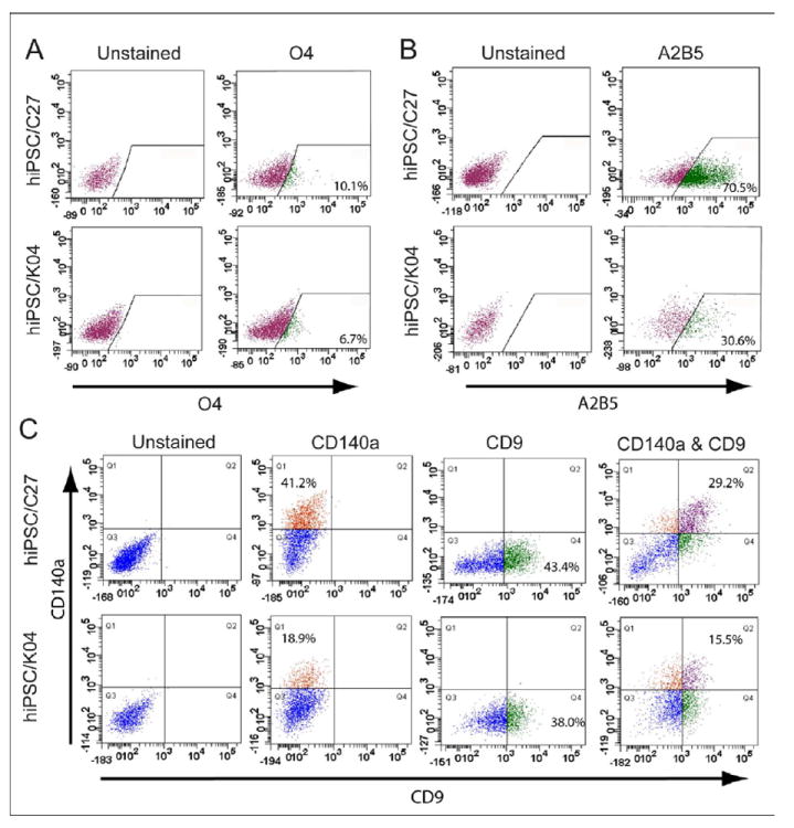
(A) hiPSC OPC-derived oligodendrocytes were recognized and isolated by fluorescence-activated cell sorting (FACS) using monoclonal antibody O4, which recognizes oligodendrocytic sulfatide. The incidence of O4+ oligodendroglia varied across different hiPSC lines, from 4 to 12% (See Supplementary Table 1; n=4-7 experiments). (B) OPCs derived from hiPSCs (C27 and K04) were readily recognized with the cell surface marker A2B5. (C) OPCs derived from either hiPSCs (C27 and K04) or hESC (WA09/H9) were readily recognized with cell surface markers, PDGFRα (CD140a) and CD9, by FACS analysis. The relative proportions of CD140a, CD9 and CD140a/CD9 double-labeled cells varied across the different cell line-derived OPCs (n=4-7 experiments).