
Figure 5. hiPSC OPCs robustly myelinate in vivo.
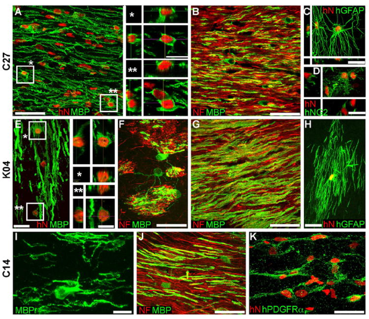
Confocal images of the callosal and capsular white matter of mice engrafted with hiPSC OPCs derived from all 3 tested hiPSC lines demonstrate dense donor-derived myelination. A-D, C27-derived; E-H, K04; I-K, C14.
A, G, and J show abundant, donor-derived myelin basic protein expression (MBP, green) by C27, K04 and C14 hiPSC OPCs (human nuclear antigen, hNA, red), respectively. Representative z-stacks of individual hNA+ oligodendrocytes are shown as asterisks in A and E. By the 19 week time-point assessed here, C27 (B), K04 (F, G), and C14 hiPSC oligodendroglia (J) robustly myelinated axons (neurofilament, NF, red). hiPSC-derived oligodendroglial morphologies exemplified in panels F (K04) and I (C14); F shows multi-axon myelination by single oligodendrocytes in the striatum.
hiPSC OPCs also generated astroglia (C, C27; H, K04), which exhibited the complex fibrous morphologies typical of human astrocytes (human-specific GFAP, green). Many cells also remained as progenitors, immunostaining for NG2 (D, C27) and human-specific PDGFαR (K, C14).
Scale: A, B, C, G, J, 50 μm; C, D, E, F, H, K, 20 μm; I, 10 μm; insets to A, E, 10 μm.