
Figure 6. hiPSC OPCs myelinate widely to greatly extend the survival of hypomyelinated mice.
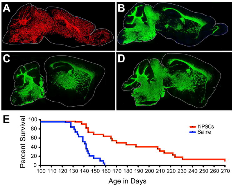
A, Dot map indicating distribution of human iPSC-derived donor cells (C27) at 7 months of age, following neonatal engraftment in a shiverer mouse brain. Widespread dispersal and chimerization by hiPSC OPCs is evident (human nuclear antigen, red). B, hiPSC OPC-derived myelination in shiverer forebrain, at 7 months; section 1 mm lateral to A. MBP-immunoreactivity (green) is all donor-derived. C, D. Myelination in sagittal sections taken at different mediolateral levels from two additional 7 month-old mice, each engrafted with C27 hiPSC OPCs at birth. E, Kaplan-Meier plot of survival of C27 iPSC-OPC implanted (n=22) vs. saline-injected (n=19) control mice. Remaining engrafted mice sacrificed for electron microscopy at 9-10 months (≥270 days).
Scale: A-B, 2 mm.
See also Figure S3.