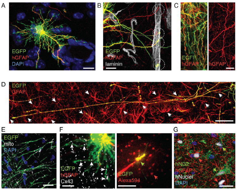
Figure 2. Human astrocytes retain hominid-specific morphology in chimeric mice.

Human protoplasmic astrocytes matured in a cell-autonomous fashion in the chimeric mouse brain environment, retaining the long GFAP+, mitochondrial-enriched processes of native human astroglia. (A) An EGFP+/hGFAP+ astrocyte makes contact with the vasculature in a one month-old chimeric mice. (B) Long, unbranched EGFP+ and hGFAP+ astrocytic processes terminated (dashed circle) on the vasculature (laminin; white) 16 days after implantation. (C) The tortuous shape of EGFP+/hGFAP+ processes in chimeric brains replicate the appearance of GFAP+ processes of interlaminar astroglia in intact human tissue. (D) An example of EGFP+/GFAP+ process that spans > 600 μm and penetrates the domains of at least 14 host murine astrocytes (white arrow), (GFAP, red). (E) Long EGFP+ processes contain a large number of mitochondria (white) in an 11 month-old chimeric mouse. (F) An EGFP+/hGFAP+ human astrocyte express C×43 (white) gap junction plaques (left panel). An EGFP+ cell (green arrow) loaded with a small gap junction permeable tracer, Alexa 594 (MW 760) in a cortical slice (P15). Alexa 594 (red) diffused into multiple neighboring EGFP- cells (red arrow). (G) Co-existence of hGFAP+ (red)/hNuclei+ (white) cells (red arrow) and hNG2+(green)/hNuclei+ cells (green arrow) in the dentate of a 12 months old chimeric mouse. (A-F): EGFP (green); (A-C): hGFAP (red).
Scale bars: 10 μm (A, C); 20 μm (B, E,G); 50 μm (D) and (F) right panel; 5 μm (F) left panel.
See also: Supplementary Figure S2.
