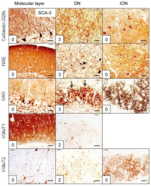
Figure 4.
The reciprocal cerebellar circuitry in SCA-3. The CAG trinucleotide repeats in this patient were 67 (expanded) and 23 (normal). Note the integrity of cerebellar molecular layer and ION. The lesion is restricted to the DN, accounting for all of the total score of 13. The arrows in GAD/DN indicate prominent grumose degeneration after visualization of GAD. Abbreviations as in fig. 1. Bars: 50 μm

