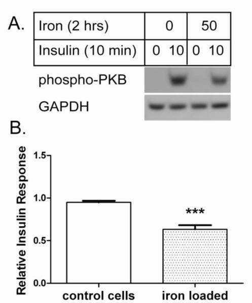
Figure 1. Iron overload decreases hepatocyte insulin response. A. Representative western blot.
Control (8-hydroxyquinoline only) and iron loaded (50 μM iron with 8-hydroxyquinoline) AML-12 cells were stimulated with 10 nM insulin and analyzed by western blot for phospho-PKB. GAPDH was analyzed as loading control. B. Relative insulin response. The insulin-stimulated phospho-PKB signals from n=18 experiments were quantified relative to GAPDH as described under Methods and normalized to control cells in each experiment; the means +/− se are plotted (*** p<0.001).

