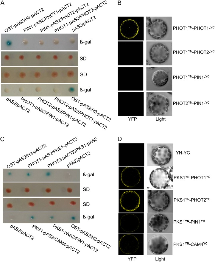
Figure 10.
PKS1 interacts with PHOT1, PHOT2, PIN1, or CAM4. A, PIN1 did not interact with PHOT1 or PHOT2, and PHOT1 also did not interact with PHOT2, in the yeast two-hybrid system. Yeast (Saccharomyces cerevisiae strain Y190) containing pAS2-PIN1 or pAS2-PHOT1 as bait and pACT2-PHOT1 or pACT2-PHOT2 as prey, or pAS2-PHOT1 or pAS2-PHOT2 as bait and pACT2-PIN1 or pACT2-PHOT1 as prey, were grown for 48 h on synthetic defined (SD) medium that lacked Trp and Leu (middle panel) and were assayed for LacZ expression by a filter-lift assay for β-galactosidase activity (β-gal; top and bottom panels). The empty vector pAS2 and pACT2 were used as negative controls. pAS2-OST1 and pACT2-H3 were used as positive controls. A blue color indicates interaction. B, PIN1 does not interact with PHOT1 or PHOT2, and PHOT1 also does not interact with PHOT2, in vivo, as determined by BiFC. Left panels show fluorescence images under confocal microscopy, and right panels show bright-field images of the cells. C, PKS1 interacted with PHOT1, PHOT2, PIN1, or CAM4 in the yeast two-hybrid system. Yeast strains containing pAS2-PHOT1 or pAS2-PKS1 as bait and pACT2-PKS1, pACT2-PHOT2, pACT2-PIN1, or pACT2-CAM4 as prey were grown for 48 h on synthetic defined medium that lacked Trp and Leu (middle panel) and were assayed for LacZ expression by a filter-lift assay for β-galactosidase activity (top and bottom panels). D, PKS1 interacted with PHOT1, PHOT2, PIN1, or CAM4 in vivo as determined by BiFC. Left panels show fluorescence images under confocal microscopy, and right panels show bright-field images of the cells. YFP, Yellow fluorescent protein; YN and YC represent pSPYNE and pSPYCE (for split YFP N-terminal/C-terminal fragment expression), respectively.

