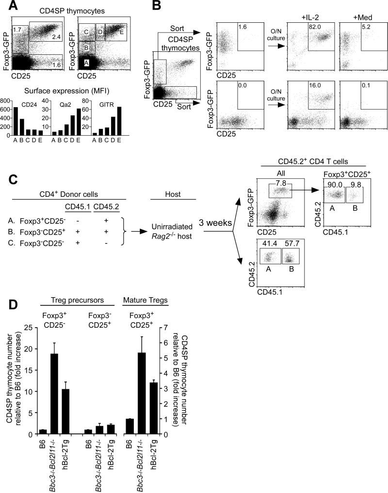
Figure 6. Foxp3+CD25- precursors of Foxp3+CD25+ mature Tregs.
(A) CD4SP thymocytes from Foxp3-GFP reporter mice (Bettelli et al., 2006) (top) and flow cytometric analysis of gated subsets (A-E) (bottom). Data are representative of 4 experiments.
(B) In vitro differentiation of sorted Foxp3+CD25− and Foxp3−CD25+ CD4SP thymocytes. Data are representative of 6 experiments.
(C) In vivo development of Foxp3+CD25+ Tregs from two Treg precursor subsets. CD4 cells were electronically sorted into subsets of Foxp3-GFP+CD25− cells (A), Foxp3-GFP−CD25+ cells (B), and Foxp3-GFP−CD25− cells (C) as indicated (left) and transferred together into unirradiated Rag2-/- host mice. Each host mouse received 3×10⁵ donor cells of A, 1×10⁵ donor cells of B, and 1×10⁶ donor cells of C. Three weeks later, host spleen and LN T cells were analyzed (right). Data are representative of 4 host mice analyzed in 2 independent experiments. (D) Effects of Puma-Bim double deficiency and hBcl-2Tg expression on precursor and mature thymic Tregs. Each CD4SP thymocyte subset was compared relative to B6 mice which was set equal to 1. Mean ± SE of 4-9 mice per group.
See also Figure S5.

