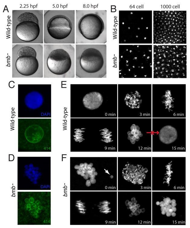
Figure 1. bmb is required for early development and proper nuclear morphology.
(A) Prior to the MBT (2.25 hpf), embryos from bmb mutant females are morphologically similar to WT embryos. After the MBT at 5.0 and 8.0 hpf, bmb mutants fail to undergo cell movements associated with epiboly and gastrulation. (B) DAPI staining of interphase nuclei during cleavage in WT (top) and bmb− (bottom) reveals multiple chromatin bodies associated with each nucleus. (C) WT and (D) bmb− interphase nuclei stained with mab414 (green) indicate that bmb− nuclei are multi-micronucleated compared to WT. (E) WT and (F) bmb− cell cycle time course at the 2 to 4 cell transition (images are projections of multiple confocal Z-slices). Top left –interphase; Top middle- prophase, Top right-metaphase, bottom left -anaphase, bottom middle- telophase, bottom right-interphase. In E and F for each time point, n=3. In this and subsequent figures ‘n’ refers to number of embryos examined (unless otherwise noted). In each case multiple nuclei or cells of each embryo were also examined.

