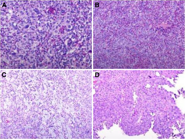
Figure 1.
Histologically, lymphoepithelioma is composed of nests, sheets and cords of undifferentiated cells with large pleomorphic nuclei and prominent nucleoli, and the cytoplasm borders are poorly defined imparting a syncytial appearance (A,B); the background consists of a conspicuous lymphoid stroma with histiocytes, neutrophils and eosinophils, the latter prominent in our patient’s case (C); the tumor is focally admixed with typical high-grade papillary urothelial carcinoma (D).

