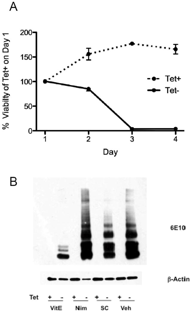
Figure 2.

A. MC65 cells were plated on Day 0 in medium with (Tet+) or without (Tet-) tetracycline. Cell viability was assessed with the MTT assay and data presented as % (SEM) of Tet+ cultures on Day 1. Tet+ cultures continued to expand over the next 2 days before reaching a plateau. In contrast, Tet- cultures showed delayed but complete toxicity that developed over Days 1 to 3. B. Cultures of MC65 cells were incubated with (Tet+) or without (Tet-) tetracylcline (1 μg/ml) in the presence of the reagents for 48 h and then harvested in RIPA buffer, proteins separated by SDS-PAGE, and then probed with antibody 6E10 that recognizes an epitope in amino acids 1–16 of Aβ peptides. All Tet+ cultures failed to display any detectable 6E10-immunorective bands. Cultures exposed to drug vehicle (Veh) show the expected multiple species of 6E10-immunoreactive bands. Incubation with α-tocopherol (VitE, 100 μM) suppressed HMW aggregates of 6E10-immunoreative species, as previously described. In contrast, incubation with nimodipine (Nim, 25 μM) or SC-51089 (SC, 50 μM) did not block the formation of HMW species. Western blot for β-actin is loading control.
