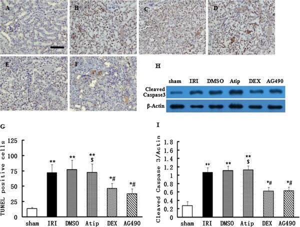
Figure 3.

The effect of dexmedetomidine inhibition on I/R-induced apoptosis of tubular epithelial cells. Representative microphotographs were taken from the kidneys of the sham (A), IRI (B), DMSO (C), Atip (D), DEX (E) and AG490 (F) groups at the time point of 48 h after renal I/R in rats. Apoptosis was evaluated by terminal deoxynucleotidyl transferase dUTP nick end (TUNEL) staining. Quantification of TUNEL positive cells was counted following renal I/R (G). Western blots for cleaved caspase 3 expression (H) of kidneys were detected after 48 h of renal I/R in all six groups. Densitometry analysis of Western blots for the ratio of cleaved caspase 3/β-Actin (I). Bar = 50 μm. Data were represented as mean ± SEM (n = 8). *P < 0.05 and **P < 0.01 vs. the sham group. #P < 0.05 vs. the IRI and DMSO groups. $P < 0.05 vs. the DEX group.
