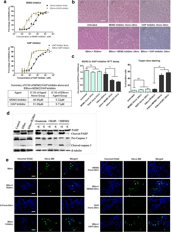
Figure 4.
MDM2 and XIAP inhibitor potentiated the cell death and caused persistent caspase activity in stressed HeLa cell. (a). Dose-dependent responsive curves of MDM2 inhibitor (Boranyl-chalcone) and XIAP inhibitor (Embelin) on unstressed cells (Inhibitor Alone) and stressed cells (E8 hrs + Inhibitor) of HeLa for 48 h treatment by MTT assay. Two-way ANOVA was used to test the significance of ethanol stress as the source of variance. IC50 of each group is summarized in table. (b). Morphology of HeLa cells recovered 24 h from stress (E8 hrs + R24 hrs) and cells treated with MDM2 inhibitor (10 μM) or XIAP inhibitor (20 μM) for 24 h during recovery period (E8hrs + inhibitor 24 hrs). Inhibitor treatment alone for 24 h was included (Scale bar: 10 μm). (c). Cell viability assay of the effects of MDM2 inhibitor (10 μM) or XIAP inhibitor (20 μM) on HeLa cells undergoing recovery from stress treatment for 24 h treatment. Left: MTT cell viability assay; Right: Trypan blue dye exclusion assay (Mean ± SEM, n = 3; ns indicates: no significance; ** indicates: p < 0.01). XIAPi stands for XIAP inhibitor; MDM2i stands for MDM2 inhibitor. (d) Western blot analysis of the effects of genistein (15 μg/ml), MDM2 inhibitor (10 μM) and XIAP inhibitor (20 μM) 24 h treatment on HeLa cells undergoing stress recovery. +E: 5.5% ethanol stress for 8 hrs followed by genistein and inhibitor treatment. -E: without ethanol stress treatment. (e). Immunostaining of cleaved caspase-3 of stressed and unstressed HeLa cells treated with genistein (15 μg/ml), MDM2 inhibitor (10 μM) and XIAP inhibitor (20 μM). Cleaved caspase-3 (Green) stained with Alexa Fluor® 488-conjugated antibody, Nuclei (Blue) stained with Hoechst 33342 (Scale bar: 100 μm).

