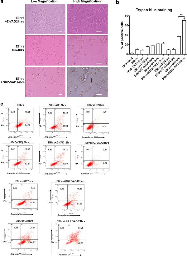
Figure 6.
Caspase activity inhibition shifted the effects of genistein to secondary necrosis. (a). Morphology of HeLa cells undergoing the recovery from stress treatment, in the presence of 100 μM Z-VAD-fmk (upper), or 15 μg/ml genistein (middle), or both (bottom) for 24 h. Arrows indicate the cell swelling and membrane rupture of shrunk cells. (b). Trypan blue dye exclusion assay of HeLa cells after the removal of stress, in the presence of 15 μg/ml genistein or 100 μM Z-VAD-fmk or both for 12 h and 24 h respectively (Data expressed as Mean ± SEM, n = 3; ** indicates: p < 0.01). (c). Annexin V and propidium iodide staining assay of HeLa cells after stress treatment and stress removal in the presence of 15 μg/ml genistein or 100 μM Z-VAD-fmk or both for 12 h and 24 h respectively. (E + Z-VAD) 8 hrs stands for ethanol stress for 8 h in the presence of Z-VAD. The percentage of events early apoptotic (lower right), and late apoptotic or necrotic (upper right) were indicated in each diagram.

