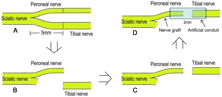
Figure 1. Surgical procedures.
(A: The right sciatic nerve and its two main branches were exposed, then the common peroneal nerve and the tibial nerve were transected at 5 mm distal to the bifurcation; B: the proximal stump of the tibial nerve and the distal stump of the common peroneal nerve were ligated and stitched to the adjacent muscle; C: the proximal stump of the common peroneal nerve and the distal stump of the tibial nerve were aligned with each other; D: the proximal stump of the common peroneal nerve served as the donor nerve and was bridged to the distal stump of the tibial nerve with an artificial conduit).

