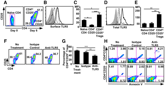
Figure 1. LR5 blockade reduced the generation of CD4hiCD25+ regulatory T cells and was independent of apoptosis.
(A) Flow cytometric analysis of the percentage of CD4hiCD25+ regulatory T cells generated on Day 6 (right panel) from naïve CD4+CD25−CD45RO− T cells (left panel). (B) Flow cytometric analysis of the expression of surface TLR5 in freshly isolated naïve CD4+CD25−CD45RO− T cells (dotted line), and CD4+CD25− (dashed line) and CD4hiCD25+ regulatory T cells (solid line) after 6 days of co-culture of naïve CD4+CD25−CD45RO− T cells with allogeneic CD40-activated B cells. Filled histogram indicates the staining obtained from isotype-matched mAb controls. (C) Mean fluorescence intensity (MFI) of the expression of surface TLR5. Data show Mean+SEM, n = 6. (D) Flow cytometric analysis of total TLR5 in freshly isolated naïve CD4+CD25−CD45RO− T cells (dotted line), CD4+CD25− (dashed line), and CD4hiCD25+ regulatory T cells (solid line) after 6 days of co-culture of naïve CD4+CD25−CD45RO− T cells with allogeneic CD40-activated B cells. Filled histogram indicates the staining obtained from isotype-matched mAb control. (E) Mean fluorescence intensity (MFI) of the expression of total TLR5. Data show Mean+SEM, n = 6. (F) Flow cytometric analysis of the generation of CD4hiCD25+ regulatory T cells with no treatment (left panel), with isotype-matched mAb (middle panel), and with anti-TLR5 blocking mAb (right panel) during the co-culture. (G) Mean percentage of CD4hiCD25+ regulatory T cells generated with no treatment, with isotype-matched mAb, and with anti-TLR5 blocking mAb. Data shown Mean+SEM, n = 6. (H) Flow cytometric analysis of the percentage of apoptotic CD4hiCD25+ regulatory T cells (upper panel) or CD4+CD25− T cells (lower panel) after 6 days of co-culture of naïve CD4+CD25−CD45RO− T cells with allogeneic CD40-activated B cells. All results shown are representative of three independent experiments. *p<0.05, **p<0.01, ***p<0.001, one way ANOVA with Tukey’s pairwise comparisons.

