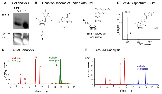
Figure 2. Reaction of BMB with tRNA following the reaction conditions described by Yang & Soell [36].
A) In-gel detection of tRNA-BMB-conjugates of total tRNA from E. coli and in-vitro-transcript (IVT) tRNA in a polyacrylamide gel. The fluorescence was imaged upon excitation at 365 nm with a GelDoc and the staining control with GelRed was imaged on a Typhoon. B) Possible reaction mechanism of BMB with uridine as an exemplary nucleoside. C) Mass spectrum, structure and main fragmentation of positively charged [M+H]+ of BMB-uridine-conjugate. The Mass transition used in (E) is indicated by an arrow. D) HPLC analysis of total tRNA E. coli reacted with BMB, digested to nucleosides and detected with a diode array detector (DAD). The red chromatogram shows nucleoside absorption at 254 nm and the green chromatogram absorption at 320 nm of BMB and its conjugates. Peaks overlapping in both chromatograms indicate possible BMB-nucleoside conjugates. E) LC-MS/MS analysis of total tRNA E. coli reacted with BMB and digested to nucleosides using the mass transitions given in Table S1 in File S1.

