
Figure 3. Sequences of 13C-NMR spectra of L. lactis cell suspensions (pH 4.8 and 5.5), showing separate resonances due to intra- and extracellular lactic acid.
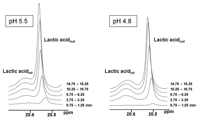
Due to the fast rate of proton exchange each resonance represents the total contribution of the two forms of lactic acid (dissociated and non-dissociated forms). The extracellular pH varies by about 0.2 units because of a lag in the addition of base in the circulating system used for the NMR experiment.