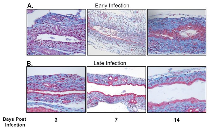
Figure 2. Histologic appearance of the extrahepatic biliary tract following infection with RRV in early versus late injected mice.
Mason’s trichrome staining of extrahepatic bile ducts after infection with RRV leads to complete obstruction of extrahepatic biliary duct in early infection group (A). In contrast, the late infection group had some mild inflammation with a patent bile duct (B). In each group, the images are magnified 40X.

