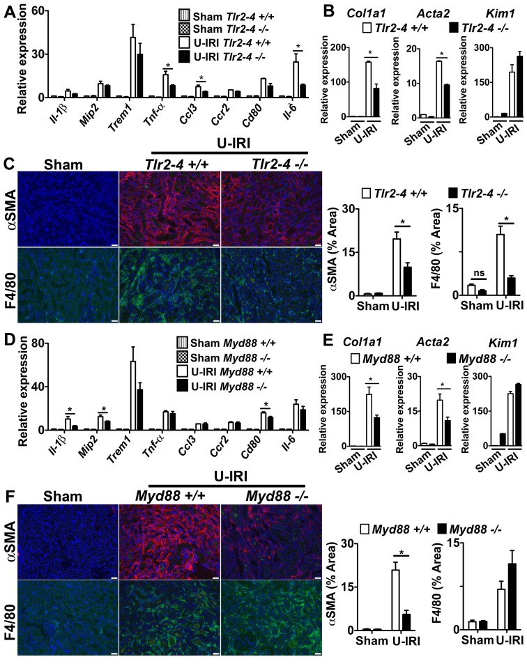
Figure 4. The TLR-2/TLR-4/MyD88 pathways play a role in fibrosis in the U-IRI model of sterile kidney injury.
(A–C) Tlr2–4−/− (D–F) Myd88−/− mice or respective controls were subjected to U-IRI and kidney harvested for tissue analysis 5 days later. Q-PCR (A,D) for different inflammatory transcripts, (B,E) pro-fibrotic transcripts, collagen1a1 (col1a1) and alpha smooth muscle actin (Acta2), and the tubule injury marker, kidney injury molecule-1 (Kim-1) from whole kidney day 5 after U-IRI. (C,F) Representative fluorescent images (left) and quantitative graphs (right) showing+αSMA (red) cells and+F4/80 cells (green). (*P<0.05, n = 5–7/group, 3 independent experiments; ns, p is not significant; Bar = 50 µm; Q-PCR results were normalized to wild type control).

