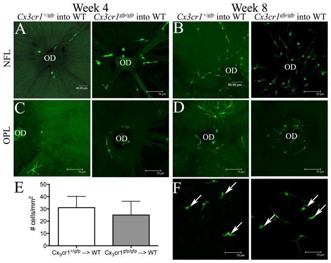
Figure 2. CX3CR1 deficiency does not influence the replenishment of retinal microglia or vitreal hyalocytes in BALB/c mice.
At 4 weeks post BM transfer, donor GFP+ cells were present in small numbers surrounding the optic disc (OD) at the level of the NFL (A), inner plexiform layer (not shown) and OPL (C) of both Cx 3 cr1 +/gfp → WT and Cx 3 cr1 gfp/gfp → WT chimeric mice. At week 8, the proportion of GFP+ cells appeared to be similar in Cx 3 cr1 +/gfp → WT (B, D; left) and Cx 3 cr1 gfp/gfp → WT chimeric mice (B, D; right). The cell density of donor GFP+ vitreal hyalocytes in Cx 3 cr1 +/gfp → WT and Cx 3 cr1 gfp/gfp → WT chimeric mice was similar at 8 weeks post BM transfer (E; P = 0.12). Examples of hyalocytes indicated by white arrows (F). n= 4 mice per group.
NFL, nerve fiber layer; OPL, outer plexiform layer.

