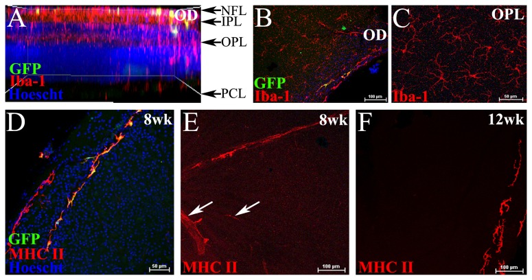
Figure 4. The acute effects of sublethal irradiation on the retina.
Confocal microscopic analysis of retinal whole mounts from C57BL/6J mice that had received sublethal irradiation (5.5 Gy) plus Cx 3 cr1 +/gfp BM transfer. Whole retinal scan viewed in side profile showing few GFP+ donor cells in the NFL and IPL of the retina at 8 weeks post-irradiation and BM transfer (A). En face image at the optic disc (OD) region showing few GFP+ cells in chimeric mice at 8 weeks (B). Host derived GFP- Iba-1+ microglia in the OPL (C). Donor derived GFP+ cells at the peripheral retina were mainly perivascular and expressed MHC Class II (D). MHC Class II was expressed on some of the inner retinal vasculature 8 weeks after sublethal irradiation (E, white arrows), but not at 12 weeks, where only perivascular cells at the periphery were MHC Class II+ (F). n= 6 mice per group.
NFL, nerve fibre layer; IPL, inner plexiform layer; OPL, outer plexiform layer; OD, optic disc; PCL, photoreceptor cell layer.

