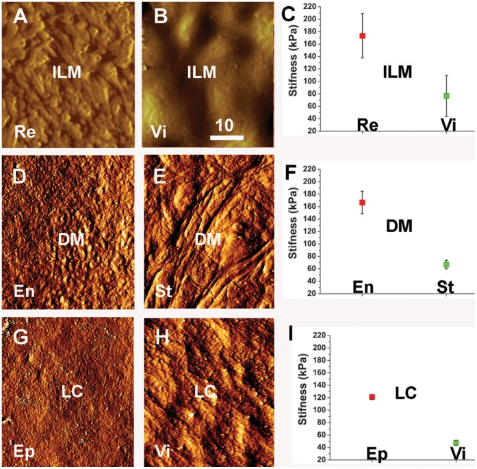
Figure 2. AFM testing of the two surfaces of the ILM (A–C), the DM (D–F) and LC (G–I).
The AFM imaging mode shows the morphological differences between the retinal (Re)/epithelial (Ep)/endothelial (En) surfaces and the vitreal (Vi)/stromal (St) surfaces of the ILM, DM and the LC. The graphs in (C, F, I) show the quantification of the stiffness measurements obtained by AFM “forced indentation”. The measurements were obtained by probing five ILMs, three DMs and three LCs. The epithelial surfaces of all tested BMs were about twice stiffer than the stromal surfaces. The differences were statistically significant. Scale bar: 10 µm.

