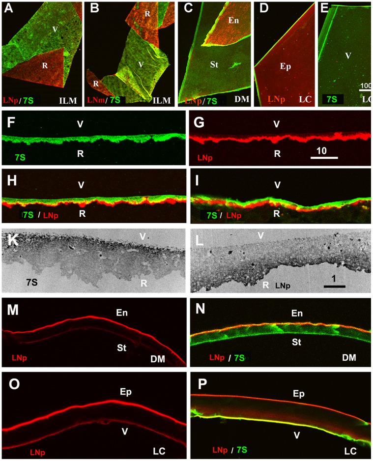
Figure 3. The asymmetric structure of human BMs as shown by double labeling of folded ILMs (A, B), folded Descemet’s membranes (DM, C) and differently mounted lens capsule segments (LC, D, E).
The retinal surface (R) of the ILM and the endothelial (En) and epithelial (Ep) sides of DM and LC were stained with a polyclonal (red, LNp; A, C, D) and a monoclonal antibody to laminin (red, LNm, B), whereas the vitreal (V) or stromal side (St) of the BMs were labeled with an antibody to the 7S domain of collagen IV α3/4/5 (green; A-C, E). The asymmetry of BMs was also detected by single and double labeling of crossections of an isolated ILM (F-H) and an ILM in situ (I). The TEM micrographs in panel (K, L) show crossections of isolated ILMs stained for 7S collagen IV α3/4/5 (K) and laminin (L). The dark label shows the localization of 7S collagen IV on the vitreal side (K) and laminin on retinal side of the ILM (L). An asymmetric distribution for laminin and collagen 7S was also detected for the Descemet’s membranes (M, N) and the lens capsule (O, P). The sections were stained for laminin (red; M-P) and collagen IV 7S (green; N, P). Scale Bars: A-E: 100 µm; F-I and M-P: 10 µm; K, L, I: 1 µm.

