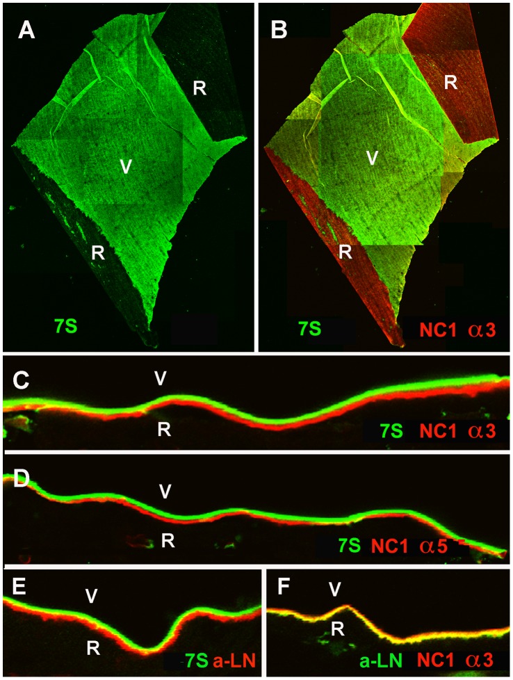
Figure 7. Distinct localization of the N and C-terminal domains of collagen IV in the human ILM.
Labeling of ILM flat mounts with an antibody specific for the 7S domain of collagen IV α3/4/5 (7S) stained only the vitreal side (V) of the BM (green, A). The epithelial/retinal side of the ILM was prominently labeled with an antibody to NC1 domain of collagen IV α3 (B, red, NC1 α3) as shown by double labeling. Double labeling of retinal crossections with antibodies to the 7S domain of collagen IV α3/4/5 (green, C-E) and the NC1 domain of collagen IV α3 (NC1 α3, red, C) or α5 (NC1 α5, red, D) confirmed the distinct distribution of the C and N-terminal domains of collagen IV in this BM. Adjacent sections were stained for 7S of collagen IV α3/4/5 (green) and laminin (LN; red; E), and laminin (red) and NC-1 collagen IV α3 (green; F). Panel (E) shows the distinct localization of laminin and the 7S domain of collagen IV, and the yellow label in panel (F) the co-localization of laminin with the NC1 domain of collagen IV. Bar: A, B: 100 µm; C-F: 10 µm.

