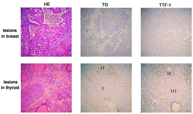
Figure 2.
Hematoxylin and eosin (HE) staining and immunostaining (TG and TTF-1) of malignant lesions from the breast and thyroid gland. Upper panel: the malignant lesion and the adjacent breast component exhibited negative staining for TG and TTF-1. Lower panel: the normal component of the thyroid tissue was positive for TG (region II) and TTF-1 (region IV); however, metastatic cancer cells (regions I and III) were negative. Magnification, ×200. TG, thyroglobulin; TTF-1, thyroid transcription factor 1.

