
Figure 3.
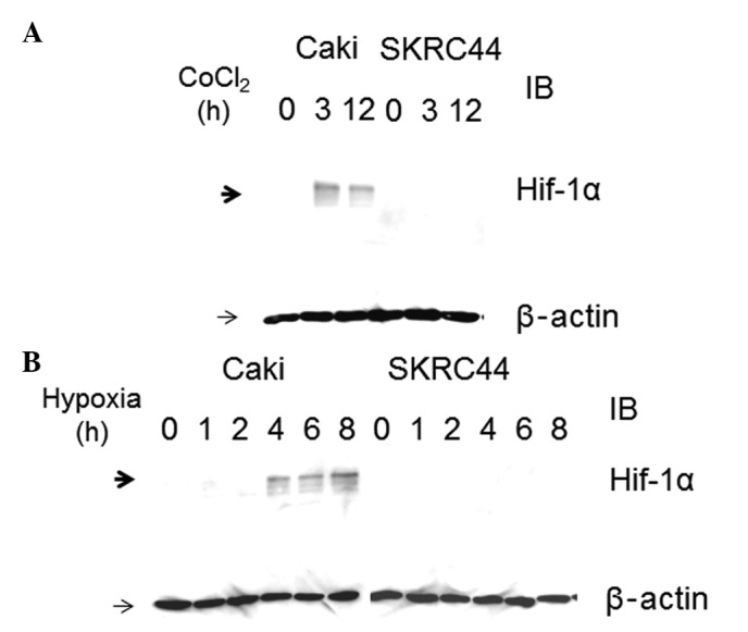
Cobalt chloride (CoCl2)- or hypoxia-induced hypoxia inducible factor (HIF)-1α expression in Caki-1 and SKRC44 cells (A) For HIF-1α induction, the cells were incubated with 125 μM CoCl2. HIF-1α and β-actin protein levels were detected by western blot analysis of whole-cell extracts, as described in the Materials and methods, and were measured at 0, 3 and 12 h subsequent to incubation. (B) Caki-1 and SKRC44 cells were exposed to hypoxia for 8 h and measured at 0, 1, 2, 4, 6 and 8 h. Hypoxia induces Hif-1α expression in Caki-1 cells, but not in SKRC44 cells. HIF-1α and β-actin protein levels were detected by western blot analysis of whole-cell extracts, as described in the Materials and methods. IB, immunoblot.