Figure 1.
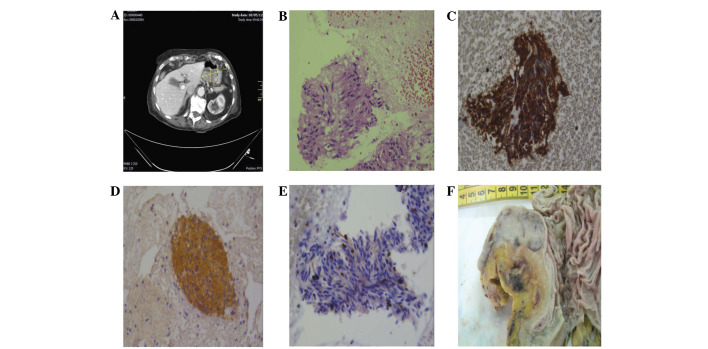
Case 1. Coronal CT scan showing a hypodense mass with not homogeneous contrast enhancement developing from the small gastric curve and causing (A) a partial reduction of the gastric lumen. (B) Cytological smears exhibiting aggregates of spindle cell elements with elongated nuclei (haematoxylin-eosin, ×160); the same elements were intensely immunoreactive for (C) vimentin (immunoperoxidase, ×200) and (D) CD117 (immunoperoxidase, ×120), showing only a sporadic nuclear immunopositivity for (E) Ki67 (immunoperoxidase, ×200). (F). The surgical specimen revealed the gastric sub-mucosal localisation of the GIST. GIST, gastrointestinal stromal tumour; CT, computed tomography.

