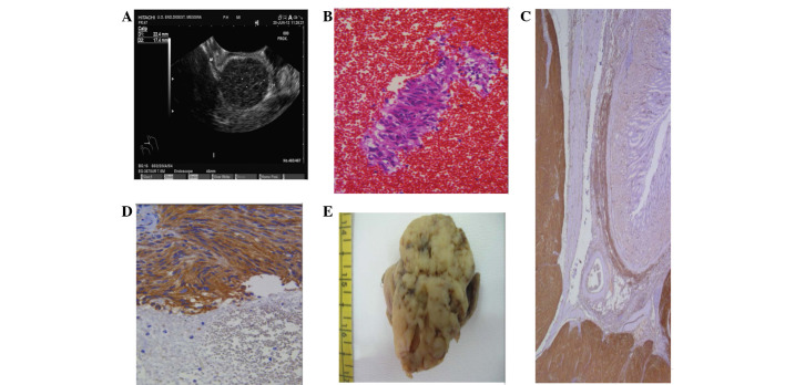
Figure 2.
Case 2. (A) EUS scanning revealed a 22.4×17.4-mm, hypoechoic, well-delimited lesion, originating from the muscle layer. (B) The cytology of the lesion was strongly suggestive of a GIST, being formed of clusters of spindle cells (immunoperoxidase staining; magnification, ×200). (C) Upon histological examination, a diffuse cytoplasmic CD117 immunoreactivity was found in the proliferative spindle cell elements of the gastric wall (immunoperoxidase staining; magnification, ×120). (D) The clusters of spindle cells were reactive for CD117, but also for CD34 (immunoperoxidase staining; magnification, ×160). (E) The cut surface of the surgical specimen showed a white-greyish nodular feature. EUS, endoscopic ultrasound; GIST, gastrointestinal stromal tumour.

