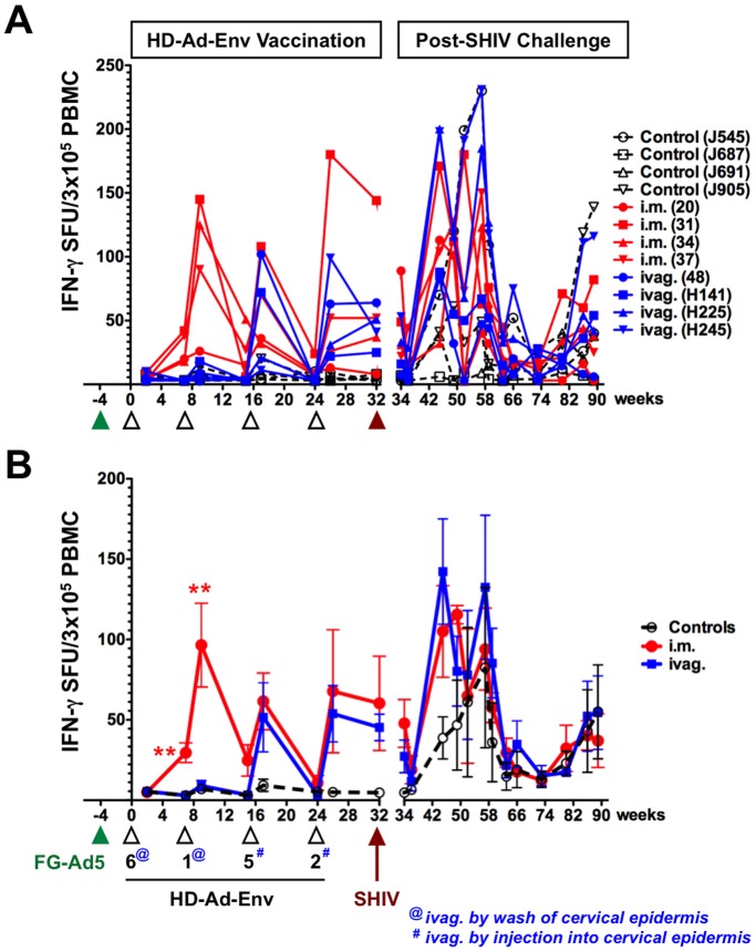
Figure 2. IFN-γ Secreting Cells from PBMCs.
PBMCs in ELISPOT plates were stimulated with 3 pools of 50 to 70 peptides spanning the gp140 region of SF162 envelope for 36 h and IFN-γ secreting spots were detected and counted. Responses in terms of IFN-γ spot forming units (SFU) for 105 total input cells were determined for individual monkeys after subtracting background values of cells cultured in the medium. A) IFN-γ secreting cells from individual animals. B) Mean IFN-γ secreting cells from each group.

