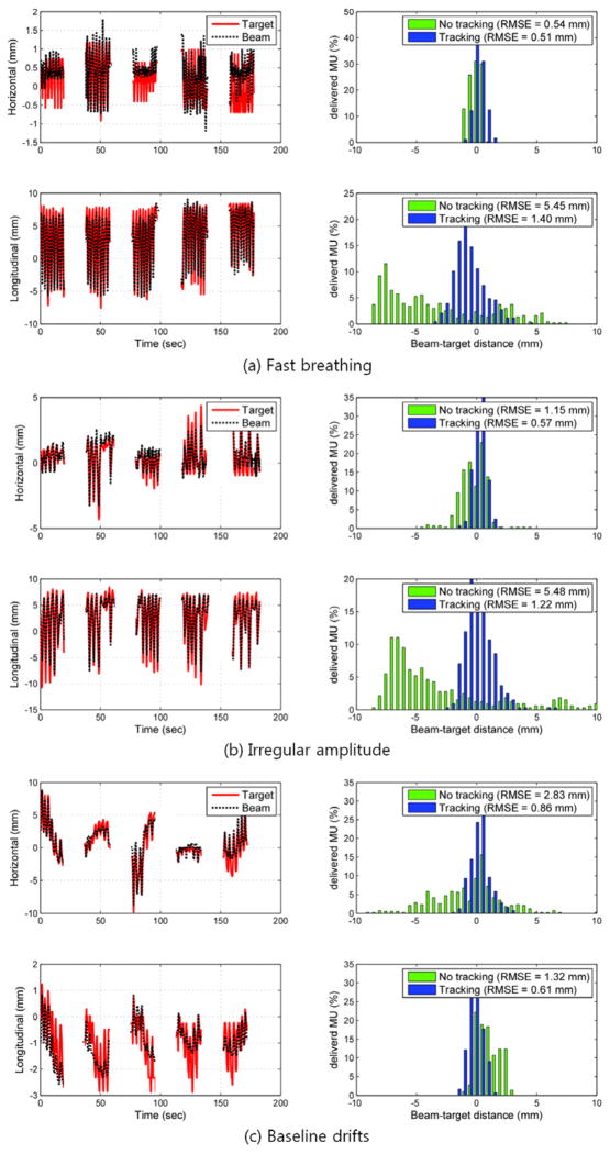
Figure 5.
The MV imager measured target position, aperture position and tracking error of three typical tumour motion traces during 5-field delivery featuring (a) fast breathing, (b) irregular amplitude, and (c) baseline drifts during the beam delivery. The left column shows the trajectory of the marker and the following MLC aperture in each direction on MV images, where the longitudinal position corresponds to motion in the SI direction. The right column shows histograms of tracking error without (no tracking) and with (tracking) real-time localisation.

