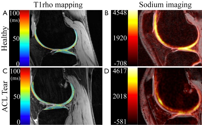
Figure 3.
Quantitative MR imaging of ACL tear knees. T1rho mapping (A,B) is applied to demonstrate the traumatic effects of ACL tear on cartilage biochemistry, compared to healthy controls. Increased heterogeneity of T1rho relaxation times within weeks of injury (B) suggests these changes occur along with the traumatic event. Sodium imaging (C,D) is also applied to reveal the impact of ACL tear on GAG content (Courtesy of Caroline Jordan, Ph.D., Stanford Dept. of Bioengineering)

