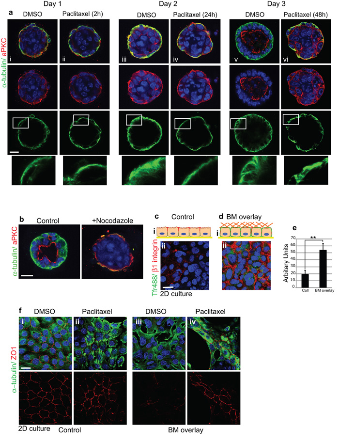
Figure 6. Dynamic MTs are required for apical relocation of aPKC and lumen formation.
(a) Time-course of polarity and lumen formation in 3D ICR acini embedded within BM-matrix and treated with paclitaxel (100 nM). Untreated acini (i, iii, v) developed apical lumens normally and MTs became orientated apico-basally. In contrast, MT stabilization (ii, iv, vi) impaired aPKC reorientation and lumen formation. Bar: 15μm.
(b) Nocodazole prevents lumen formation.
(ci) Schematic Z-view of polarized MECs in monolayer with apical tight junctions and basolateral integrins. (ii) Integrins are absent from top surface (extracellular domain antibody to β1; red) and apical membrane cannot internalize Tfr-488 from the media.
(d) MECs overlaid with BM matrix display β1-integrins at the top (i) schematic Z-view (ii) confocal view and TfR-488 internalization from the media. Bar: 8μm.
(e) Tfr488 uptake was quantified using a fluorescence plate reader. Histogram shows a single experiment representative of n=3. **p < 0.02; ***p< 0.05.
(f) Confluent monolayers of ICR MECs displaying apical tight junctions (i) were treated with DMSO (i, iii) or with paclitaxel (ii, iv) for 1h prior to BM-overlay (iii, iv). BM-overlay induced ZO1 tight junction disruption in DMSO treated cells but paclitaxel treatment prevented the disruption. Bar: 8μm.

