Figure 3.
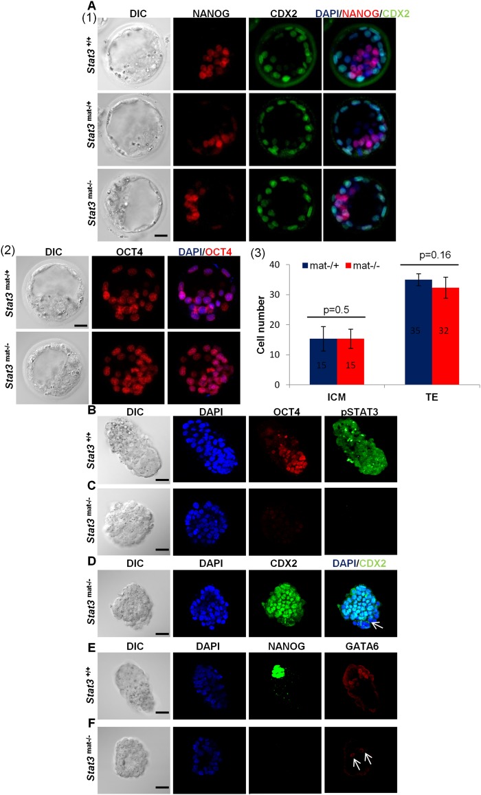
Maternal and paternal Stat3 knockout embryos isolated at E4.5 exhibit loss of the EPI and PE lineages but not TE. (A) (Panel 1) Immunodetection of NANOG and CDX2 in Stat3+/+, Stat3mat−/+, and Stat3mat−/− early blastocysts isolated at E3.5. (Panel 2) Immunodetection of OCT4 in Stat3mat−/+ and Stat3mat/− E3.5 embryos. (Panel 3) Bar graphs showing the average cell number in ICM and TE lineages of Stat3mat−/+ (n = 3) and Stat3mat−/− (n = 3) early blastocysts isolated at E3.5. ICM and TE cells are identified as NANOG+/CDX2− and CDX2+ cells, respectively. (B,C) Immunodetection of pSTAT3 and OCT4 in Stat3+/+ and Stat3mat/− E4.5 embryos. (D) Immunodetection of CDX2 in Stat3mat−/− E4.5 embryos. White arrow indicates the presence of CDX2-negative cells in the knockout embryos. (E,F) Immunodetection of NANOG and GATA6 in Stat3+/+ and Stat3mat−/− E4.5 embryos. White arrows indicate the presence of remaining GATA6-positive cells in the knockout embryos. Bar, 20 μm.

