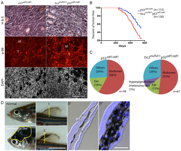
Fig. 2.
Increased mTORC1 signaling, enhanced tumorigenesis and altered tumor spectrum from the addition of a tsc2 heterozygous allele to a p53zdf1/zdf1 background. (A) H&E, phospho-S6 and DAPI (nuclei) staining of abdominal tumors from p53zdf1/zdf1 and tsc2;p53 compound mutant zebrafish. Left panels: invasive region of a p53zdf1/zdf1 tumor; right panels: tsc2;p53 compound mutant tumor. Circular structures are skeletal muscle bundles encircled by invasive tumor cells (pink in the H&E staining, but dark areas in the immunofluorescence images). Phospho-S6 signal is red and DAPI is white. M, muscle; T, tumor. Scale bar: 100 μm. (B) Kaplan-Meier tumor-free survival. Blue line is p53zdf1/zdf1 zebrafish, red line is tsc2;p53 compound mutant zebrafish. 50% of p53zdf1/zdf1 zebrafish had tumors at ∼550 days of life compared with 450 days for tsc2;p53 compound mutant zebrafish. n=115 and 130, respectively. This reduction in latency to tumor detection was statistically significant using the Log-rank test (P<0.0001). (C) Spectrum of tumor types from p53zdf1/zdf1 and tsc2;p53 compound mutant zebrafish. Tumor types were broadly similar with the exception of melanoma-like tumors, which were seen only in tsc2;p53 compound mutant zebrafish. (D) Normal zebrafish head and dorsal fin in the top panels and melanoma-like tumors on the head and dorsal fin in the bottom panels. Dorsal fins were sectioned at the region marked ‘1’ (normal) and ‘2’ (tumor) and stained with DAPI in D′ and D″, respectively. Scale bars: 5 mm (D); 100 μm (D″).

