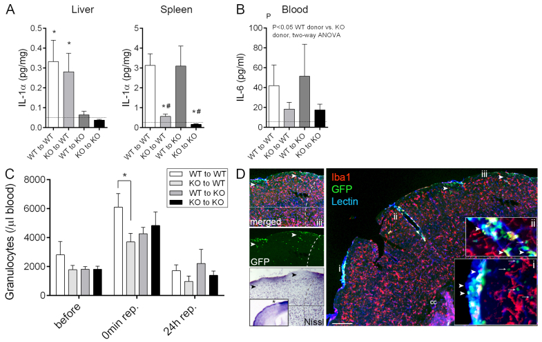
Fig. 2.
Lack of haematopoietic-derived IL-1 results in altered systemic inflammatory responses after cerebral ischaemia. (A) IL-1α production in the liver is dependent on host-derived cells with slow turnover, whereas splenic leukocytes expressing IL-1α are readily replaced by bone-marrow-derived cells within 6-8 weeks after bone marrow transplantation. IL-1α levels in liver and spleen homogenates at 24 hours reperfusion are shown. (B) Plasma IL-6 levels are reduced 24 hours after MCAo in mice receiving IL-1αβ KO bone marrow. Dashed lines in panels A and B indicate the detection limit of the assay. (C) Stroke results in a rapid increase in circulating granulocytes (P<0.01, 45 minutes after the onset of ischaemia compared with prior to occlusion), which is blunted in WT mice transplanted with IL-1αβ KO bone marrow. (D) Recruitment of donor-derived (GFP+, green) leukocytes (arrowheads) to the brain 24 hours after MCAo is most pronounced in the meninges (lectin, blue) and in large cortical blood vessels (lectin, blue). Tissue infiltration of blood-borne cells is seen only in superficial cortical layers (green, arrows) and is minimal deep in the brain parenchyma, with very minor contribution of donor-derived (GFP+) macrophages (Iba1, red). Inserts show a higher magnification of the areas labelled with i and ii. (iii) Recruitment of blood-borne cells into the brain (GFP, arrowheads) is associated with neuronal loss (Nissl, asterisk on insert is showing the area of interest in the ipsilateral hemisphere). Dashed line indicates the boundary of the infarct. Cc, corpus callosum. Scale bar: 200 μm. *P<0.05 versus KO to WT, and KO to KO, respectively (A, left), *P<0.05 versus WT to WT, and #P<0.05 versus WT to KO (A, right), oneway ANOVA followed by Tukey’s multiple post-hoc comparison (A), two-way ANOVA (B), and two-way ANOVA followed by Bonferroni’s multiple comparison (C). n=7–9, data are expressed as mean ± s.e.m.

