
Figure 2.
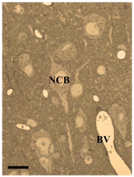
LM photomicrograph of unstained 0.5 micron semi-thin section of epoxy embedded mouse cortex shows crisp cellular details and good contrast.
NCB Neuronal cell body, BV blood vessel. Scale bar 10 μm. Animal use in this experiment was conducted in strict accordance to our institutional animal care and use committee guidelines.