
Figure 1.
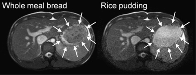
Representative example of axial MRI images of the abdomen of a healthy volunteer fed on one occasion WMB meal (left) and RP meal (right) on another occasion, taken at t=0. The closed arrowheads indicate the body of the stomach. In this balanced gradient echo MRI sequence, liquid appears bright and viscous solutions appear dark. The images show that the WMB meal formed a dark bolus occupying most of the distal stomach, surrounded by bright fluid at the edges, close to the stomach walls. The RP meal rapidly separated into an upper brighter fluid liquid and a lower particulate phase in the stomach.