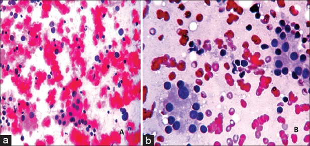
Figure 1.

(a) Microphotograph showing Hashimoto's thyroiditis with thyroid follicular cells showing Hurthle cell change and lymphocytes in the background, categorized into the benign category (Pap, ×400), (b) Microphotograph of a smear categorized as AFLUS, showing predominantly benign thyroid follicular cells in sheets, with some cells showing anisonucleosis and forming microfollicles (Pap, ×400)
