Figure 1.
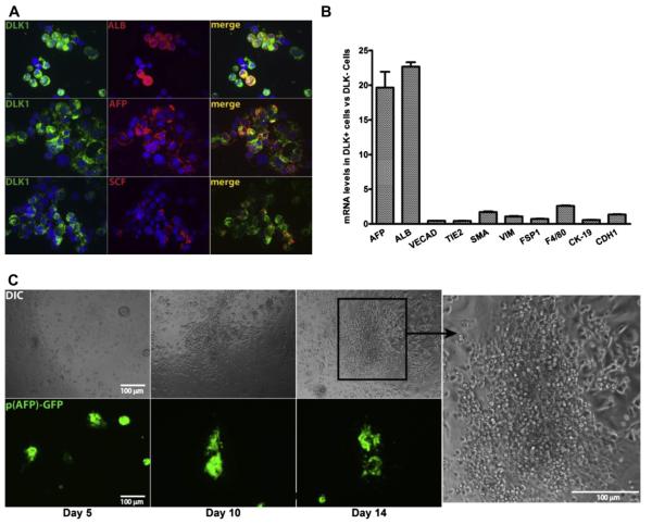
Purified DLK+ fetal hepatic progenitors support the growth of hematopoietic cells in ex vivo culture. (A) Double immunocytochemistry for DLK (green) and ALB, AFP, or SCF (red) expression in fetal liver DLK+ cells purified by magnetic beads. Only a fraction of DLK+ cells express SCF and ALB, but almost every DLK+ cell is also AFP+. (B) Relative enrichment of mRNAs encoding markers for hepatic progenitors (AFP, ALB), endothelial cells (VECAD, TIE2), mesenchymal cells (α-SMA, VIM, FSP1), Kupffer cells (F4/80) and bile duct epithelial cells (CK-19, CDH1) in purified DLK+ versus DLK− cells. The mRNA level of each gene was determined by qPCR and normalized against ribosomal RNA. The ratio of mRNA levels in DLK+ cells versus DLK− cells is plotted (n 5 3 with SEM). (C) Hematopoietic cells contaminating the DLK+ cells expanded adjacent to fetal hepatic cells in serum containing medium; 25,000 DLK+ cells purified from Tg (AFP-GFP) mice were cultured in one well of a 96-well plate in IMDM medium plus 10% serum. The survival of fetal hepatic progenitors in ex vivo culture was tracked by their expression of GFP (bottom panels). Clusters of small, round hematopoieitc cells start to grow adjacent to GFP+ hepatic cells at day 10 and continue to expand through day 14 (Differential Interference Contrast microscopy; top panels).

