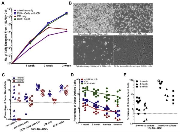
Figure 3.
Fetal DLK+ hepatic progenitors support long-term expansion of HSCs. The coculture experiment shown in Figures 2E–2G was extended to 3 weeks. (A) Numbers of cells generated from one SLAM+ HSC after 1, 2, and 3 weeks of culture with DLK+ cells plus CM, with DLK+ cells alone, with CM alone, or with cytokines only. (B) Representative phase contrast micrographs of the progeny of 50–100 SLAM+ HSCs cultured with DLK+ cells, CM, or cytokines alone for 2 weeks in one well of a six-well plate. Also shown are DLK+ cells cultured alone with no HSCs added (bottom right). (C) Scatter plots showing the percentage of donor-derived CD45.1+ peripheral blood cells in mice transplanted with the progeny of 10 SLAM+ HSCs after a 2-week culture (n = 7–8). (D) The percentage of donor-derived blood cells in mice at successive times after transplantation with the progeny of 10 SLAM+ HSCs cultured with DLK+ cells, CM, or cytokines only. The data for months 2 and 4 after transplantation are the same as in (C). The average percentage repopulation of each group of recipient mice at the indicated times after transplantation was calculated and shown as trend lines. DLK+ cells, but not CM, expanded long-term HSCs that maintain their repopulating ability after 6 months. (E) Three-week coculture further expanded HSCs. Comparison of the percentage reconstitution from the progeny of one SLAM+ cell after a 2-week culture (n = 8) with those after 3 weeks of coculture (n = 5). One mouse transplanted with cells expanded from 3 weeks of coculture died because of anemia between 1 and 2 months after transplantation (open circle).

